The more effective the diagnostics that doctors have at their disposal, the better armed they are to to diagnose and treat breast cancer. Two new cancer tools—breast seed localization and 3D mammography—being employed at Capital Health’s Center for Comprehensive Breast Care will help doctors do just that. Here’s what they are and why you might want to consider them for yourself.
BREAST SEED LOCALIZATION
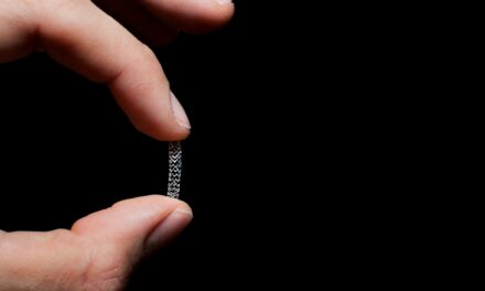
What is it: Breast seed localization is a procedure in which a radioactive iodine seed is inserted into the breast at the site of the abnormal tissue to help your surgeon find the area during surgery when it is too small to be seen or felt by hand. Seed localization enables radiologists to pinpoint areas of concern, so your surgeon can remove the abnormal tissue with as little disturbance to surrounding healthy breast tissue as possible.
When did Capital Health start using it? August 2015
How does it work: The radiologist will numb the area with a local anesthetic, insert the needle containing the radioactive seed, leave the seed in position, and take a mammogram image to make sure the seed is properly placed. “The radiologist can put the seed in up to 5 days in advance, and then the seed is removed at the time of surgery. Precautions are taken in the operating room to ensure the entire seed is removed,” explains Lisa Allen, M.D., director of the Center for Comprehensive Breast Care.
l What’s good about it: “The seed allows us to localize benign and malignant tumors without the discomfort of having a needle and/or wire remain in the breast prior to surgery (needle localization).” Dr. Allen says. “It is the same cost to the patient and has been shown to reduce the amount of tissue removed in patients with benign tumors.”
3D MAMMOGRAPHY
What is it: While 2D mammography typically takes x-rays of the patient’s breast from two angles, 3D mammography, also known as tomosynthesis, takes multiple images of the breast along a short arc, and it takes just a few seconds to complete the study. A computer then assembles the images to produce clear, highly focused 3D images throughout the breast.
When did Capital Health start using it? May 2015
How does it work: “The 3D allows us to see through the breast tissue because we are looking at a series of 1mm slices, instead of an overlapping image,” says Anne Moch, M.D., a radiologist at Capital Health.
What’s good about it: “In my personal experience, there have been several cases where the 3D mammography has helped me determine whether it’s overlapping tissue, so we don’t have to bring a patient back,” Dr. Moch says. “On the other side of the spectrum, we had one case where a young patient had very dense breasts. We were scrolling through the 3D images, and we saw a cancer—there was a mass.” Supporting Dr. Moch’s anecdotal experience, studies show an increased cancer detection rate of about 25 percent among all breast cancers and a decreased recall rate of about 15 percent.


